Sanodent Clinique Radiology Center
At Sanodent Clinique, we prioritize precise, fast, and safe diagnosis. That is why our clinic is equipped with a modern radiology center, fully outfitted to perform all necessary dental imaging investigations in one place.
We use state-of-the-art technology from Planmeca, internationally recognized for its quality, accuracy, and safety. Our equipment is designed to provide high-resolution images with minimal radiation exposure, including dedicated programs for children.
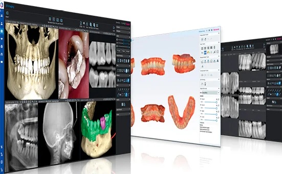
All imaging investigations are digitally stored on a secure server, allowing our doctors to access the patient’s imaging history at any time for monitoring progress and optimizing treatment planning.
3D Computed Tomography (CBCT)
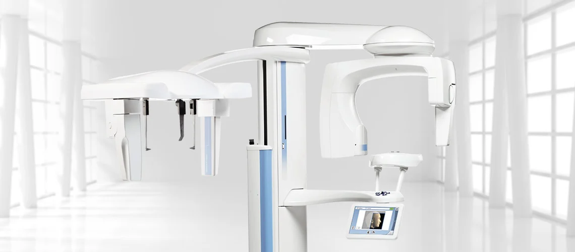
3D computed tomography represents the modern standard for detailed diagnosis of dento-maxillary structures. At our center, we use the Planmeca ProMax® 3D Classic, a high-performance scanner equipped with an integrated cephalostat.
This technology provides highly precise three-dimensional images, essential for:
-
Dental implantology
-
Oral surgery
-
Orthodontics
-
Evaluation of impacted teeth or deep infections
Advantages of 3D tomography at our clinic:
-
Detailed 3D volumetric images
-
Reduced radiation exposure thanks to advanced Planmeca technology
-
Special pediatric mode to minimize radiation dose
-
Fast diagnosis and predictable treatment planning
2D Panoramic X-ray (OPG)
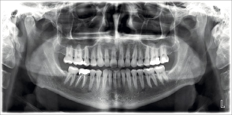
Panoramic radiography (OPG) provides an overall image of the entire dentition, jawbones, and temporomandibular joints.
It is used for:
-
General evaluations
-
Detection of hidden cavities
-
Identification of infections or cysts
-
Planning complex dental treatments
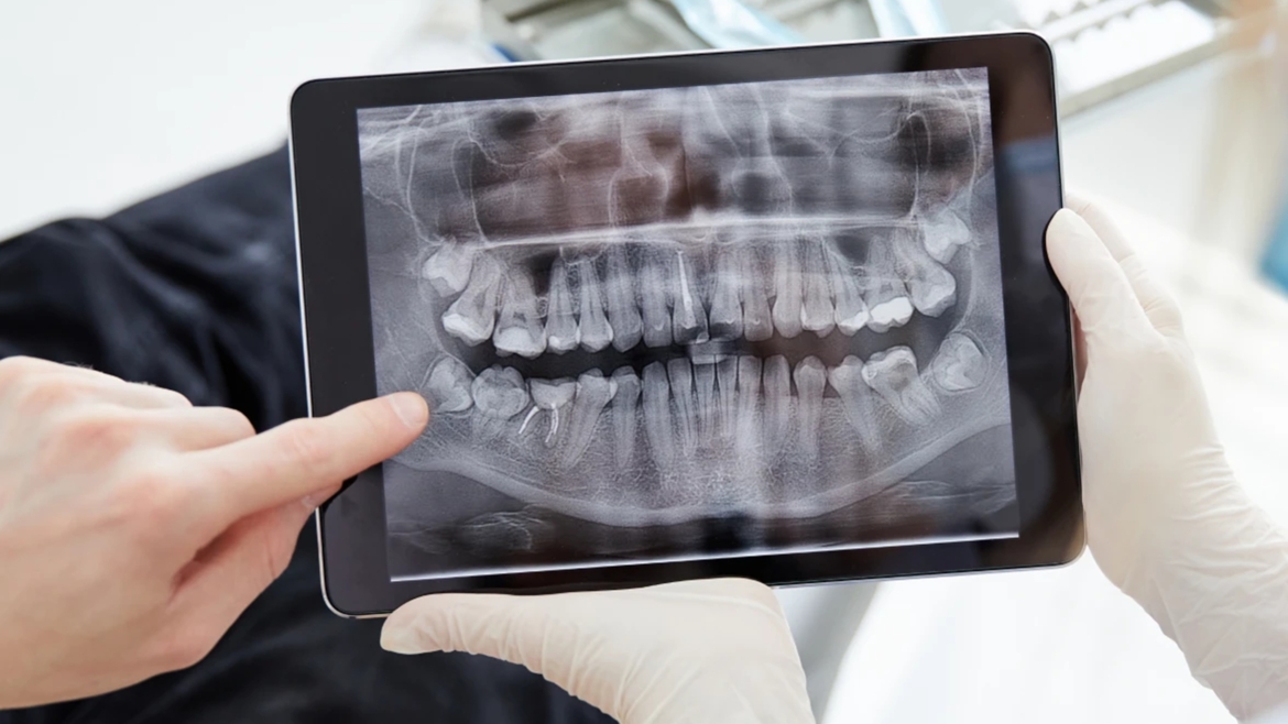
The procedure is quick, comfortable, and involves minimal radiation exposure, making it suitable for both adults and children.
Cephalometric X-ray (Teleradiography) for Orthodontics
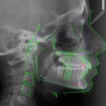
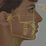
Teleradiography is essential for planning orthodontic treatments. Thanks to the integrated cephalostat in our Planmeca ProMax® 3D Classic system, we perform precise cephalometric radiographs necessary for analyzing jaw and tooth positioning.
This allows the orthodontist to:
-
Establish an accurate diagnosis
-
Analyze the facial profile
-
Plan orthodontic treatment
-
Monitor progress over time
The result is a personalized and efficient treatment plan tailored to each patient.
Digital Intraoral Radiography (Visiography)
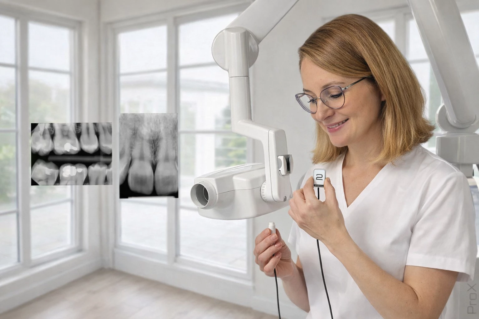
For detailed investigations of a single tooth or a specific area, we use the Planmeca ProX™ digital imaging system.
Visiography is essential for:
-
Root canal treatments (endodontics)
-
Detection of early-stage cavities
-
Verification of dental restorations
Benefits of digital visiography:
-
Very low radiation exposure
-
Instant images displayed on screen
-
High diagnostic accuracy
-
Increased patient comfort
Safety, Precision, and Digital Monitoring